Our products
HISTOLOGY
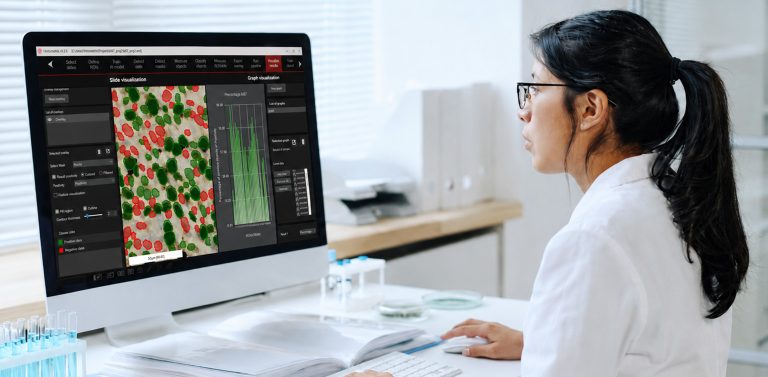
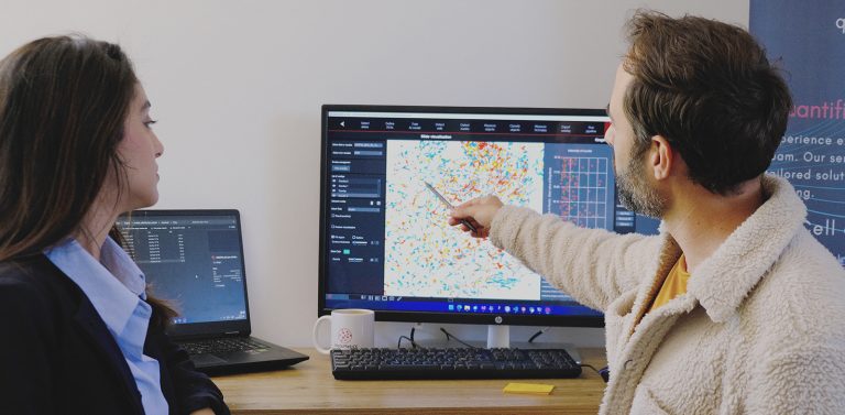
HistoMetriX software
HistoMetrix is a powerful and intuitive software designed specifically for biologists and pathologists. You can easily annotate precise regions, and our advanced deep-learning technologies automate the detection and quantification of tissue structures, and cells, saving you time and improving accuracy.
HISTOLOGY
Histological Image Analysis Services
- Customized software solutions with ergonomics UIX
- Artificial intelligence for automatic analysis
- Industrialization of analyses in laboratories
3D ORGANOID
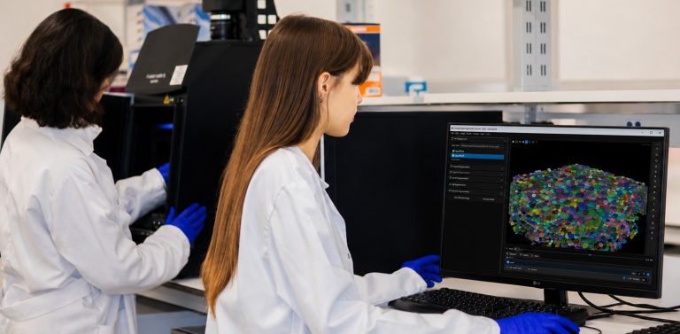
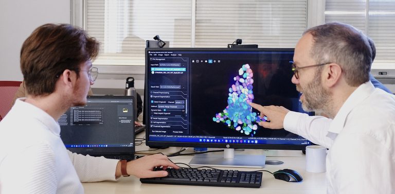
AssayScope Software
AssayScope is a user-friendly application that enables you to visualize and manipulate images of 3D cell culture, spheroids and organoids. With AssayScope, you can gain a deeper understanding of the complex 3D structures of tissues, helping you to identify the most promising molecules and treatments.
3D ORGANOID
3D Organoid Image Analysis Services
- Multi-scale segmentation: nuclei, cells, and organoids
- Extraction of morphological and topological features
- High-content screening (HCS) powered by AI

Who is it for?
Quantacell develops advanced image analysis workflows for histology, multiplex imaging, spatial biology and 3D cellular models, enabling quantitative characterization of tissues, organoids and complex biological systems.
-
Oncology
Quantitative analysis of tumor tissues and cancer organoids to characterize tumor architecture and microenvironment.
(Ki67+ cell quantification, tumor–stroma segmentation, immune infiltration analysis, spatial cell interaction metrics) -
Neuroscience
Image analysis of neuronal cultures and brain organoids to study neuronal morphology and network organization.
(neurite length quantification, synapse density measurement, neuronal cell counting, network morphology analysis) -
Dermatology & Dermocosmetics
Quantitative evaluation of reconstructed skin models and histological sections to measure tissue structure and treatment effects.
(epidermal thickness measurement, collagen density quantification, keratinocyte proliferation analysis) -
Immunology & Inflammation
Spatial analysis of immune cell populations and tissue microenvironments using multiplex and histological imaging.
(immune cell detection, spatial distribution analysis, cell neighborhood analysis, biomarker expression quantification) -
Toxicology & Drug Screening
Quantitative assessment of compound effects in cellular systems and organoids.
(cell viability quantification, apoptosis detection, morphological profiling, organoid size and structure measurement)

Our partners
“QUANTACELL is a human-scale company capable of tackling ambitious scientific challenges by combining technical expertise, a strong understanding of medical issues, and close collaboration with clinical teams.”
— Prof. Vanessa Lacheretz Szablewski, Professor of Anatomopathology, Montpellier University Hospital.
“Our collaboration with QuantaCell helped accelerate our 3D organoid analyses and supported the generation of scientific results that led to several publications in leading international journals. This work subsequently enabled the launch of new projects and international collaborations, and our approaches were praised by experts in the field, as was the unique expertise developed by QuantaCell.”
— Dr. Anne Beghin, Research Assistant Professor & Facility Manager, Microscopy Core Facility, Mechanobiology Institute, National University of Singapore
“Thanks to the HistoMetriX software, my team was able to easily extract advanced quantifications from pulmonary fibrosis images, greatly accelerating data analysis in our ANR project.”
— Chloé Féral, Team Leader, IRCAN
“Using HistoMetriX enabled me to make rapid progress in my PhD project, particularly by precisely quantifying experimental biases and improving the analysis of head and neck tumors.”
— Dr. Camille Dovin, Doctor of Anatomopathology, Montpellier University Hospital
“AssayScope provides advanced capabilities for 3D organoid analysis via multiple segmentation pipelines that include, among other features, spatial information on nuclear volume, cell volume, and various other shape descriptors. They offer timely technical support via both e-mail and virtual calls that greatly facilitate the integration of AssayScope into our research workflow.”
— Dr Guilherme Nader, Assistant Professor of Pathology and Laboratory Medicine, Perelman School of Medicine, University of Pennsylvania
Publications