Study of the directionality of axon growth
Automatic detection of the growth of axons in microscopy of fluorescence
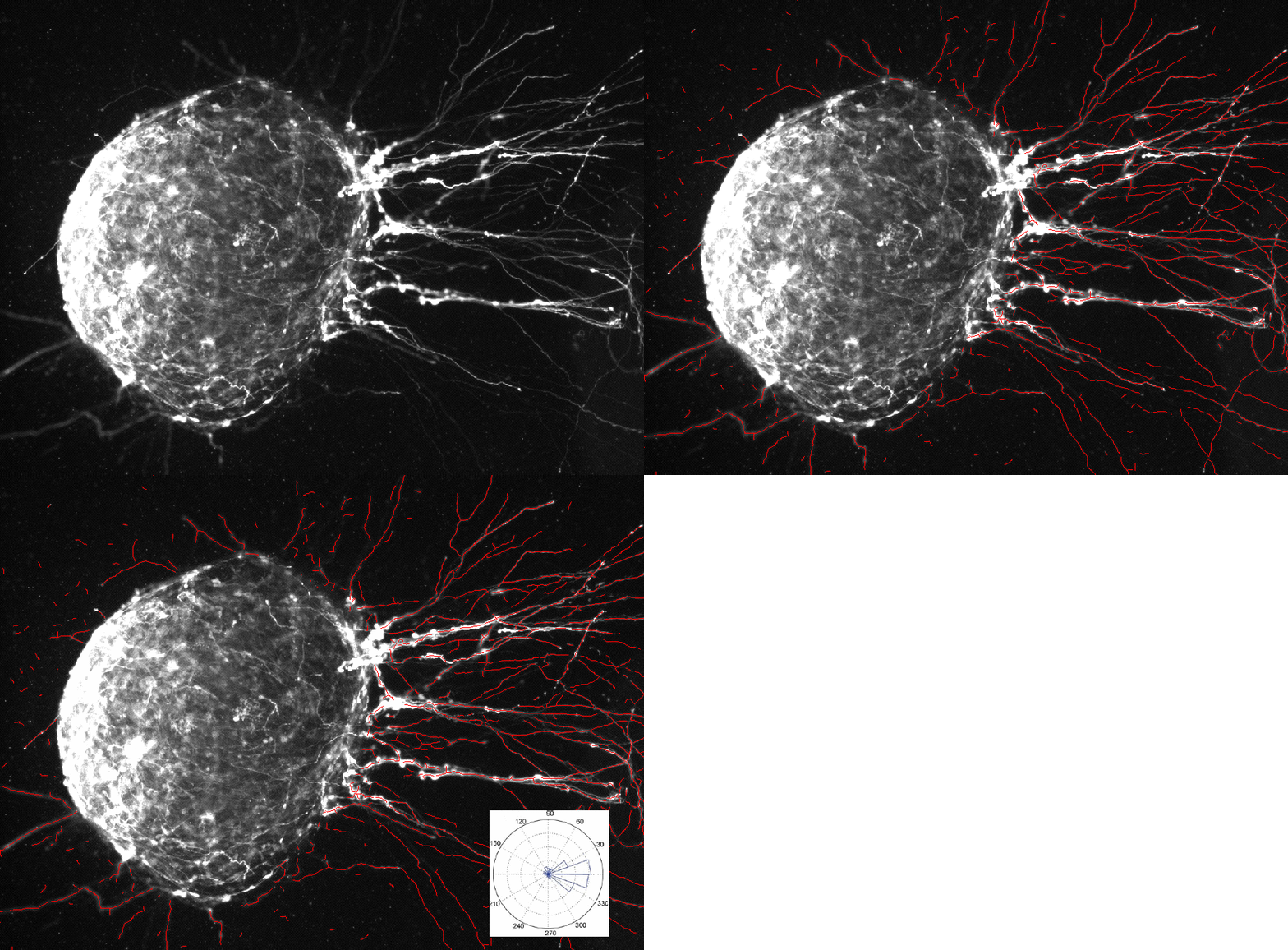
An explant of the medulla is cultivated in the presence of a chemical attractor. Directionality of the axon growth is studied. In the following illustration, the explant is labeled in GFP alpha-tubulin. Axons outside of the explant are automatically detected. Their direction is represented in the rose diagram, showing the influence of the chemical attractor, in a statistical, reproducible and unbiased manner.

Credit : Séverine Marcos and Evelyne Bloch-Gallego (Institut Cochin, Paris, France).
References
Tubulin Tyrosination Is Required for the Proper Organization and Pathfinding of the Growth Cone. Séverine Marcos, Julie Moreau, Stéphanie Backer, Didier Job, Annie Andrieux, Evelyne Bloch-Gallego, PLoS ONE 4, 4 (2009) e5405.
Interested in softwares or services?

Software for histological samples and TMA
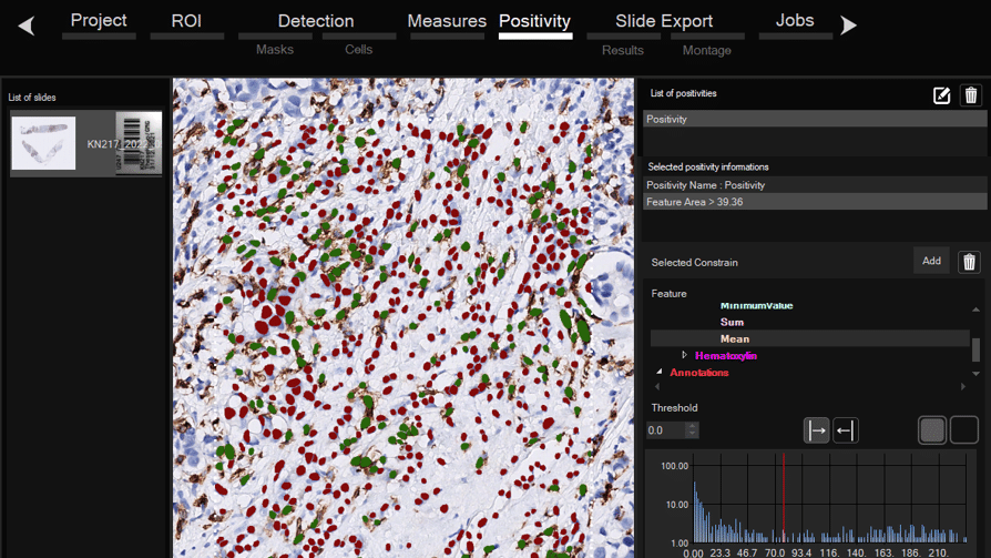
Automatically detects and quantifies your histological slides easily, without requiring bioinformatics expertise. IHC/Fluorescence/Deep learning

Software for organoid, spheroid and 3D cell culture and tissue
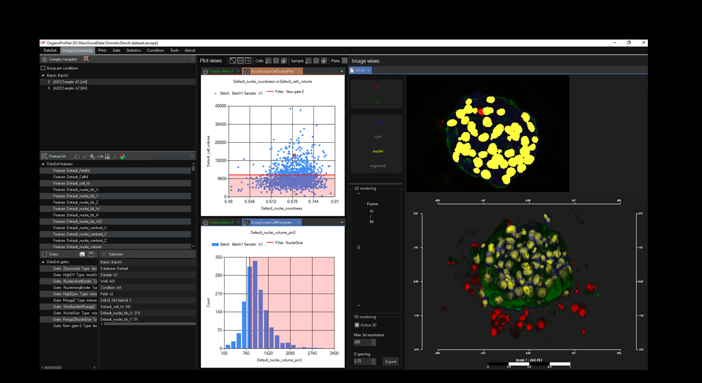
Automatic 3D segmentation and high-content screening analysis software to navigate your assays and monitor drug effects

Services of analysis and characterization
- Image analysis in patients studies
- Characterization of drug/compounds effects
- Automatic detection of rare events

Software development for biomedical imaging
- Customized software solutions with ergonomics UIX
- AI for automatic analysis
- Industrialization of analyses in laboratories
QuantaCell, Campus Balard, IHU
Contact
+33 (0) 9 83 33 81 90
300 av. du Pr Emile Jeanbrau
34090 Montpellier, France