Analysis of plant structure by electron microscopy
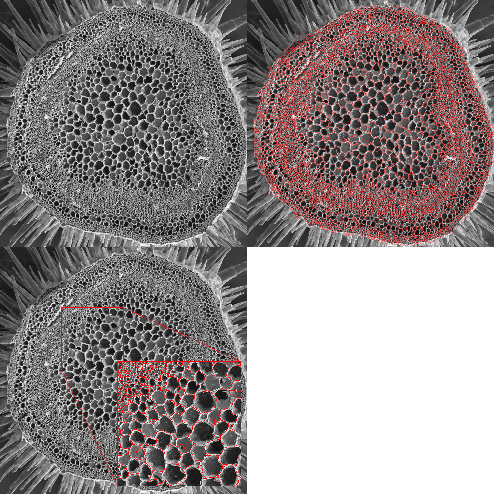
Scanning electron microscopy (SEM) images are ideal for image analysis and automatic quantification.

The following example shows a section of a winged tobacco seed (Nicotiana alata) or a large number of vascular bundles. Thanks to automatic analysis, 6250 bundles were detected and counted.
Crédits (CC pd) Louisa Howard from Dartmouth College EM facility
Interested in softwares or services?

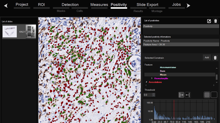
Software for histological samples and TMA
Automatically detects and quantifies your histological slides easily, without requiring bioinformatics expertise. IHC/Fluorescence/Deep learning

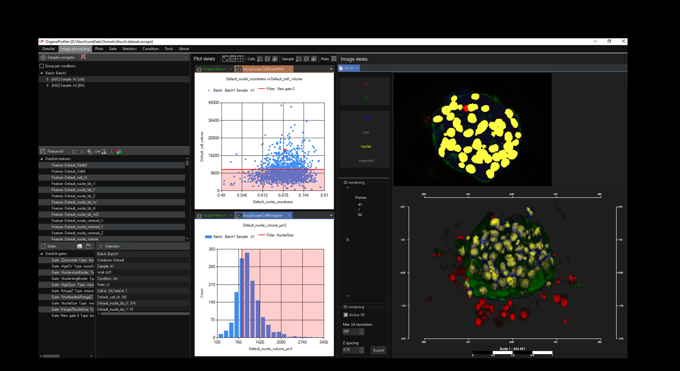
Software for organoid, spheroid and 3D cell culture and tissue
Automatic 3D segmentation and high-content screening analysis software to navigate your assays and monitor drug effects

Services of analysis and characterization
- Image analysis in patients studies
- Characterization of drug/compounds effects
- Automatic detection of rare events

Software development for biomedical imaging
- Customized software solutions with ergonomics UIX
- AI for automatic analysis
- Industrialization of analyses in laboratories
QuantaCell, Hôpital Saint Eloi, IRMB
Contact
+33 (0) 9 83 33 81 90
80 av Augustin Fliche
34090 Montpellier, France