Mixed segmentation of neurons and astroglia
Analysis of brain sliced labeled in fluorescence
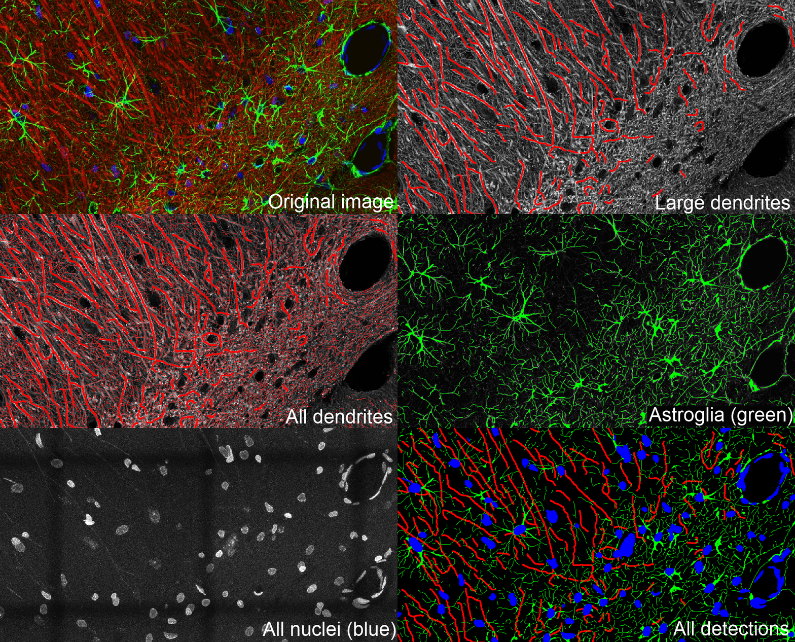
This confocal image (Leica SP5 63x) represents multipolar neurons (red) and astroglial cells (green).

An advanced segmentation is applied to every channel of the image. Neuron dendrites (labeled by denditric microtubules) are detected with a first segmentation of thick dendrites. This segmentation is completed by the detection of fine dendrites. Astrocytes are segmented in the green channel and the nuclei in the blue. Segmentation of various channels allows an analysis of fine colocalization between astrocytes and neurites.
Credits : Christopher Wallace, Ginger Withers et Tony Cooke (CC Attribution Only).
Interested in softwares or services?

Software for histological samples and TMA
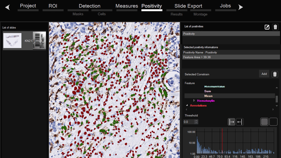
Automatically detects and quantifies your histological slides easily, without requiring bioinformatics expertise. IHC/Fluorescence/Deep learning

Software for organoid, spheroid and 3D cell culture and tissue
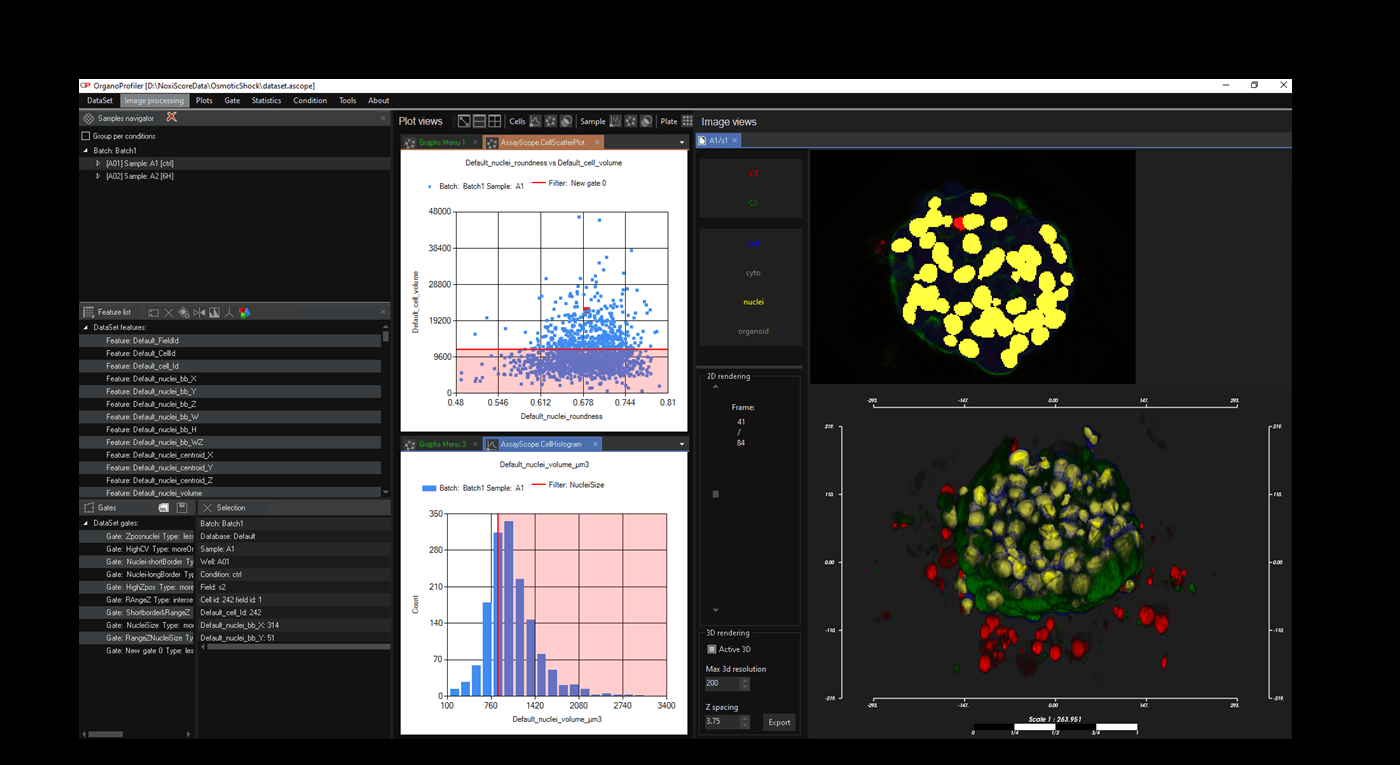
Automatic 3D segmentation and high-content screening analysis software to navigate your assays and monitor drug effects

Services of analysis and characterization
- Image analysis in patients studies
- Characterization of drug/compounds effects
- Automatic detection of rare events

Software development for biomedical imaging
- Customized software solutions with ergonomics UIX
- AI for automatic analysis
- Industrialization of analyses in laboratories
QuantaCell, Hôpital Saint Eloi, IRMB
Contact
+33 (0) 9 83 33 81 90
80 av Augustin Fliche
34090 Montpellier, France