Monitoring organ morphogenesis through time
Morphological analysis of mammary glands and the network of lactation
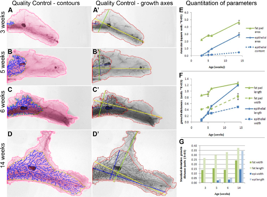
In this example the morphological transformations of the mammary gland are analyzed in adult mouse during the lactation phases. The mammary glands are extracted and plated into slides. The acquisition is made on macroscopes with a standard camera.

Quantification of the parameters dedicated to organ morphology
Possibly morphological measurements made on organ are numerous :
- Detection of the fat pad (red) with measurement of the width and the height
- Detection of the mammary epithelium (yellow) with measurement of the width and the height
- Detection of the network of lobular epithelium (blue)
- Analysis of the network of the lobular epithelium to determine the number of endings and connections
- Detection of the lymph node
These measurements allow to monitor the transformations of the mammary glands from 3 to 15 weeks mice. The parameters are illustrated in graphs.
References
Development of MammoQuant: An Automated Quantitative Tool for Standardized Image Analysis of Murine Mammary Gland Morphogenesis. Law, Yan Nei; Racine, Victor; Ang, Pei Ling; Mohamed, Hanifa; Soo, Piang Chin; Veltmaat, Jacqueline M.; Lee, HweeKuan. Journal of Medical Imaging and Health Informatics, Volume 2, Number 4, December 2012 , pp. 352-365(14)
QuantaCell, Hôpital Saint Eloi, IRMB
Contact
+33 (0) 9 83 33 81 90
80 av Augustin Fliche
34090 Montpellier, France