Tissue analysis
The automatic quantification of tissues serves to reproduce the interpretation of pathologists, by means of algorithms. The tissue sections are arranged on microscope slides and digitized with a slide scanner.
Analysis of plant structure by electron microscopy
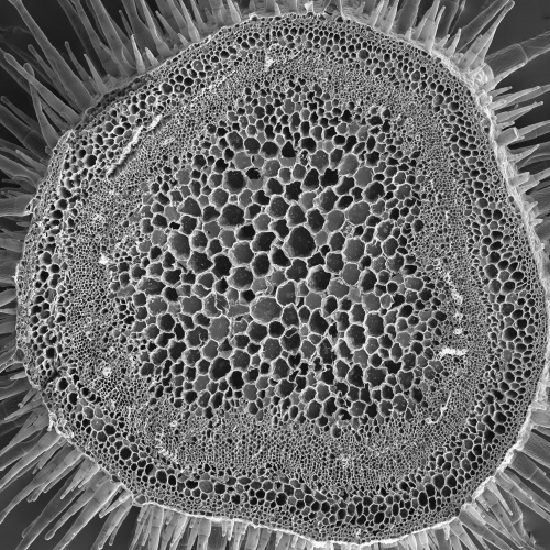
Scanning electron microscopy (SEM) images are ideal for image analysis and automatic quantification
Time-lapse tracking of organelles
The measurement of the dynamics of cell compartments is essential to the understanding of their mechanisms. It goes through the acquisition fast 2D or 3D images to be compatible with object tracking in time. The trackSpatio-temporal analysis of the cell compartments
Merging heterogeneous information
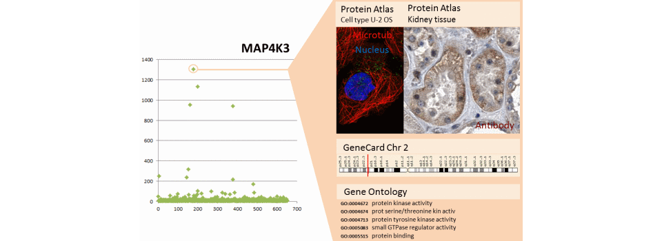
The mass of information on the biological and chemical data accessible via the Internet is huge. This information can be used to enrich the experimental results.
Study of the directionality of axon growth
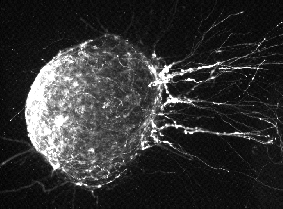
An explant of the medulla is cultivated in the presence of a chemical attractor. Directionality of the axon growth is studied.
Morphological analysis of tissues
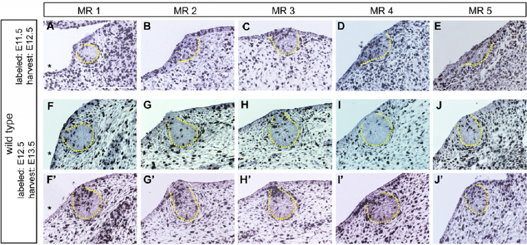
The use of the image analysis in developmental biology allows to monitor morphological parameters through the development of organs and tissues. The following examples illustrate for mice the development of mammary glands in the embryonic phase and to the adult.
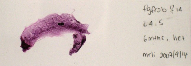
Monitoring organ morphogenesis through time
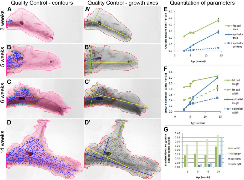
In this example the morphological transformations of the mammary gland are analyzed in adult mouse during the lactation phases. The mammary glands are extracted and plated into slides. The acquisition is made on macroscopes with a standard camera.
Detection of tissue structures
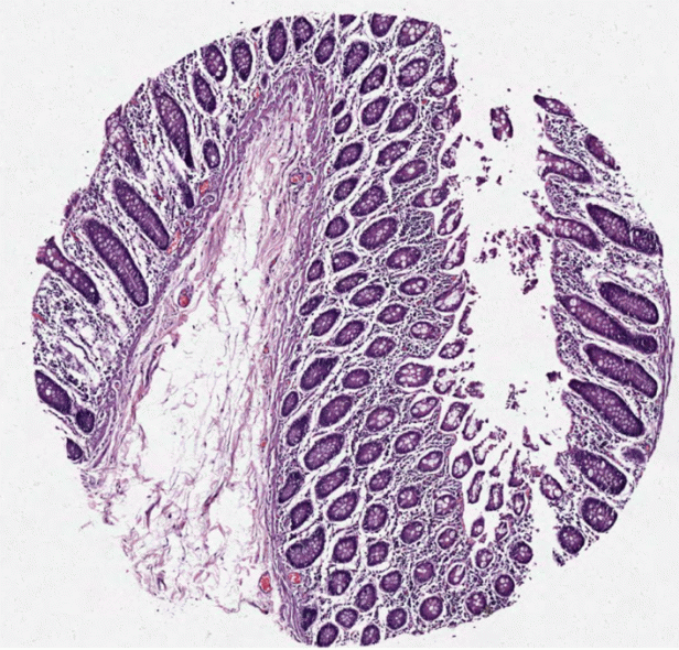
Tissue analysis needs a stage of segmentation which consists in separating the various tissular structures. In the following example, a segmentation is applied to small intestine section.
Deep learning for low contrast objects
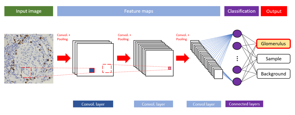
What is Deep Learning?
Deep Learning is a part of machine learning methods. It’s a technique enabling computers to learn human’s skills.
Analysis of 3D colocalization
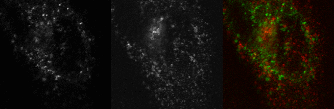
The colocalization is an important tool for cell biologists. This information allows to measure the promiscuity between two fluorescent molecules in the cell.